Home » Without Label » Back Bones Diagram - Back Bones Diagram / Divisions Of The Skeletal System ... / At the back of each bone in the spine (vertebra) are bony points called processes, which muscles attach to.
Back Bones Diagram - Back Bones Diagram / Divisions Of The Skeletal System ... / At the back of each bone in the spine (vertebra) are bony points called processes, which muscles attach to.
Back Bones Diagram - Back Bones Diagram / Divisions Of The Skeletal System ... / At the back of each bone in the spine (vertebra) are bony points called processes, which muscles attach to.. Muscle or tendon injuries can occur anywhere in the body. It also covers some common conditions and injuries that can affect the back. Each lumbar spinal level is numbered from top to bottom—l1 through l5, or l6. Exercises can strengthen the core muscles that support the spine and. Our latest youtube film is ready to run.
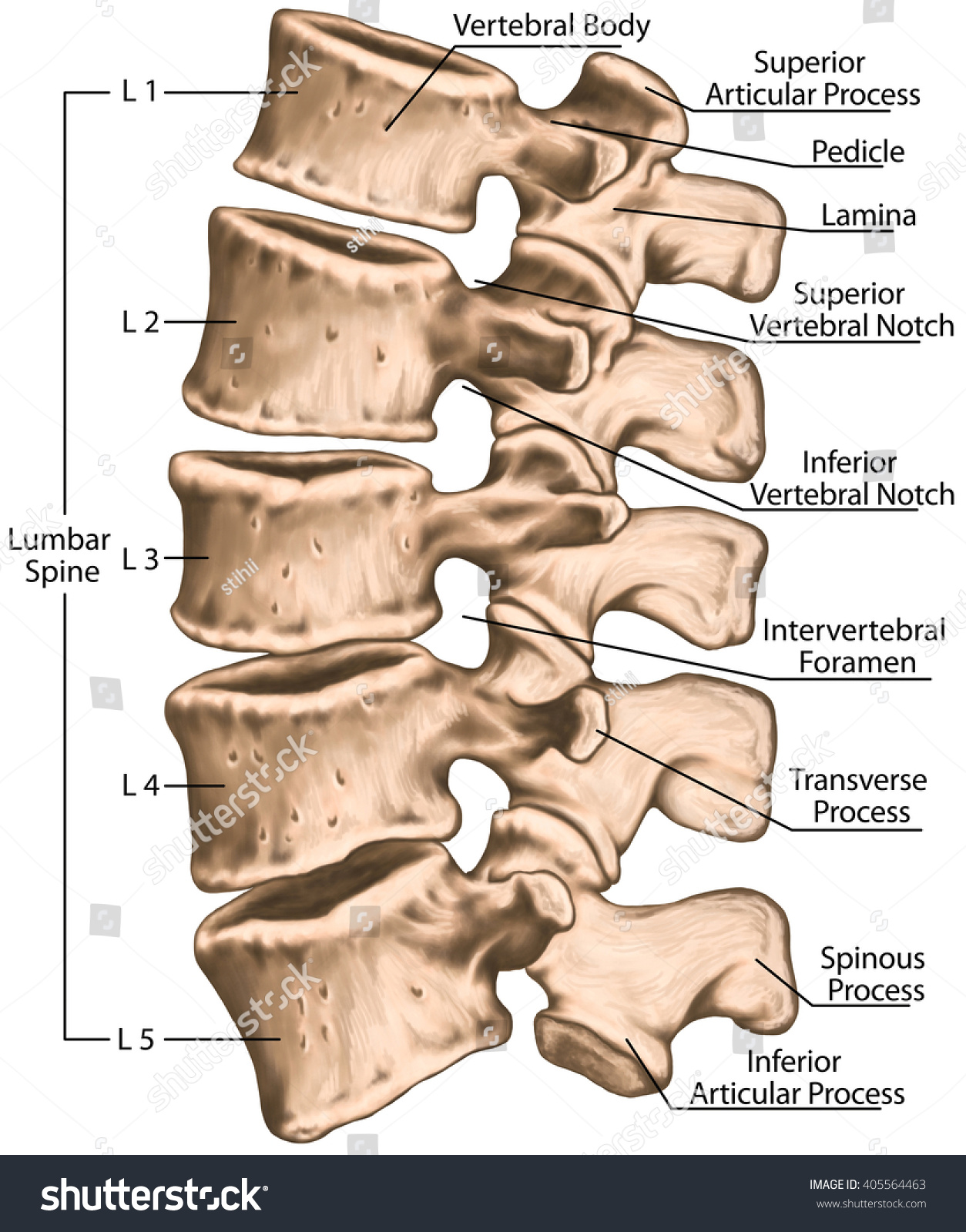
The sacrum is made of five fused bones, of which the s1 is the topmost. The vertebrae, which stack like spools of thread, support the back and protect the spinal cord. The spine anatomy is a complex structure. Add to cart add to cart add to cart add to cart customer rating: Spinal anatomy and back pain.
Back Bones Diagram / Vintage Anatomy Skeleton Images - The ... from image.shutterstock.com The atlas is a ring of bone made up of two lateral masses joined at. Your lower back contains 5 vertebral bones stacked above each other with intervertebral discs in between. In the back and elsewhere in the body, tendons attach muscles to bones. So many patients receive diagnostic imaging reports full of terms and anatomical locations which are unknown and mysterious to them. Human body poster 24 x 36in. At the back of each bone in the spine (vertebra) are bony points called processes, which muscles attach to. It is designed to be incredibly strong, protecting the highly sensitive nerve roots, yet highly flexible, providing for mobility on many different planes. At the same time the bones grow larger by growing back into the growth plates.
Each lumbar spinal level is numbered from top to bottom—l1 through l5, or l6.
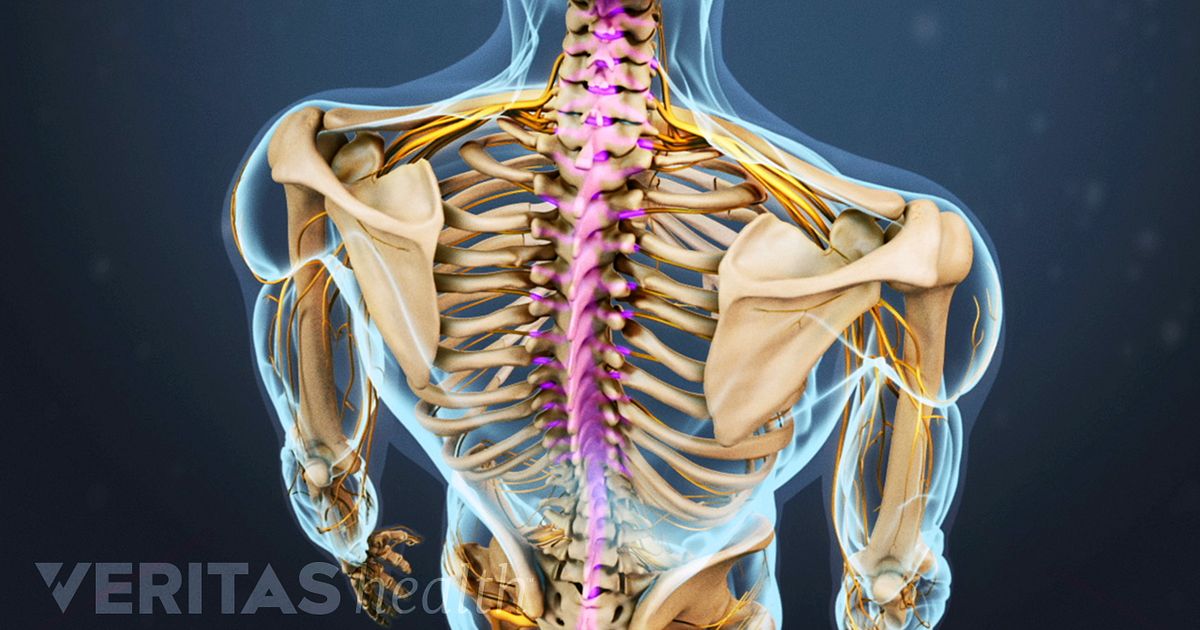
Bone diagram forehead (frontal bone) nose bones (nasals) cheek bone (zygoma) upper jaw (maxilla) lower jaw (mandible) breast bone (sternum) upper arm bone. The vertebrae, which stack like spools of thread, support the back and protect the spinal cord. Key parts of your spine include vertebrae (bones), disks, nerves and the spinal cord. Spinal vertebrae bone spine vertebra toracica spinal cord spine structure back diagram spine sections spinal cord vertebrae spinal structure health diagram. The disks that cushion vertebrae may compress with age or injury, leading to a herniated disk. There are three parts to the trapezius. The column can be divided into five different regions, with each region characterised by a different vertebral structure. Atlas (c1) the atlas is the first cervical vertebra and therefore abbreviated c1. The lumbar spine is the lower back that begins below the last thoracic vertebra (t12) and ends at the top of the sacral spine, or sacrum (s1). The occiput (co), also known as the occipital bone, is a flat bone that forms the back of the head. We are pleased to provide you with the picture named anatomy of back muscles diagram.we hope this picture anatomy of back muscles diagram can help you study and research. Each typical vertebra consists of a body, an arch and three processes that stem from. The lumbar spine connects to the thoracic spine above and the hips below.
The occiput (co), also known as the occipital bone, is a flat bone that forms the back of the head. Can you feel the bumps of your vertebrae along your back? The lower part of the trapezius ascends and depresses the scapula, while the transverse or middle region of the trapezius is what retracts the. The neck (cervical) and low back (lumbar) regions have a slight concave curve, and the thoracic and sacral regions have a gentle convex curve (fig. At the same time the bones grow larger by growing back into the growth plates.
Spinal Cord Stimulation: The Trial Period from embed.widencdn.net The disks that cushion vertebrae may compress with age or injury, leading to a herniated disk. It is also known as the vertebral column. These bones are connected at the back with specialized joints. The vast difference in height and limb length between birth and adulthood are mainly the result of endochondral ossification in the. Human body poster 24 x 36in. Individual anatomical structures include 2: For more anatomy content please follow us and visit our website: The lumbosacral joint is the joint that connects these bones.
Just need a glimpse, leave your valuable advice let us know , and subscribe us!
Bones of the pelvis and lower back. The neck (cervical) and low back (lumbar) regions have a slight concave curve, and the thoracic and sacral regions have a gentle convex curve (fig. This process continues until the end of puberty, when the growth plate stops growing and the bones fuse permanently into a single bone. Related posts of human back bones diagram bone structure birds. Exercises can strengthen the core muscles that support the spine and. The spine or backbone consists of 26 small bones or vertebrae. The first seven bones (vertebrae) of your spine form your neck. Individual anatomical structures include 2: These bones are connected at the back with specialized joints. It is designed to be incredibly strong, protecting the highly sensitive nerve roots, yet highly flexible, providing for mobility on many different planes. Seven cervical vertebrae in the neck, twelve thoracic vertebrae in the torso and five lumbar vertebrae in the lower back. Female anatomy human body classroom educational chart cool wall decor art print poster 24x36. The spine runs from the base of your skull down the length of your back, going all the way down to your pelvis.
Human body poster 24 x 36in. There are three parts to the trapezius. Bone diagram forehead (frontal bone) nose bones (nasals) cheek bone (zygoma) upper jaw (maxilla) lower jaw (mandible) breast bone (sternum) upper arm bone. 5.0 out of 5 stars: Just need a glimpse, leave your valuable advice let us know , and subscribe us!
35 Diagram Of Back Bones - Wiring Diagram Database from o.quizlet.com The sacrum is made of five fused bones, of which the s1 is the topmost. The column can be divided into five different regions, with each region characterised by a different vertebral structure. In the back and elsewhere in the body, tendons attach muscles to bones. It is also known as the vertebral column. The vast difference in height and limb length between birth and adulthood are mainly the result of endochondral ossification in the. A tough, springy disc of cartilage sits between the vertebrae of your spine. Bones, discs, and joints in your lower back. When most people mention their back, what they are actually referring to is their spine.
The first seven bones (vertebrae) of your spine form your neck.
Vertebrae are the structural constituents of the spine.there are 33 vertebrae in total; The column can be divided into five different regions, with each region characterised by a different vertebral structure. Spinal anatomy is a remarkable combination of strong bones, flexible ligaments and tendons, large muscles and highly sensitive nerves. The spine runs from the base of your skull down the length of your back, going all the way down to your pelvis. The spine diagram the spine diagram shown below, consists of many bones or vertebrae,soft discs,the spinal cord, and spinal nerves. A tough, springy disc of cartilage sits between the vertebrae of your spine. The spine or backbone consists of 26 small bones or vertebrae. Can you feel the bumps of your vertebrae along your back? Each lumbar spinal level is numbered from top to bottom—l1 through l5, or l6. This article looks at the anatomy of the back, including bones, muscles, and nerves. In the back and elsewhere in the body, tendons attach muscles to bones. Lateral labeled diagram of the human vertebral spinal column showing vertebrae numbering order and the 5 different regions of the spine. Spinal anatomy and back pain.